1. Research filtration characteristics of reservoir rocks:
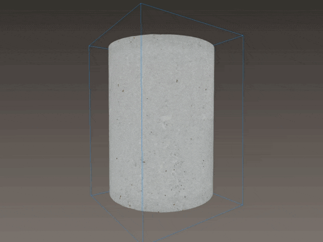
Channels formed by acid treatment of carbonates
Distribution of different size pores by volume of rock
2. Visualization of the filtration experiments in the X-ray tomograph cabinet:
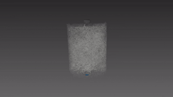
Visualization of water filtration process through the sample under 100 kPa
3. Paleontological researches of remains and archeological artifacts by non-destructive method:
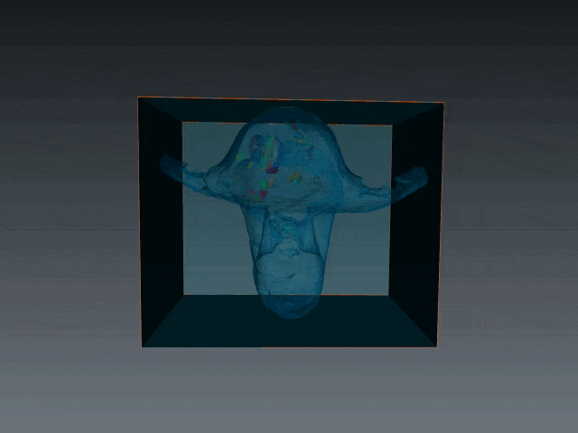
Shell form and inner structure of Nautilus
4. Defectoscopy of industrial products:
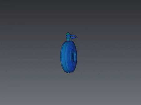
Diagnostic of the pocket watch mechanism
Peculiarities of X-Ray tomographic imaging:
- Max. sample size: width (diameter) – 230 mm, height – 420 mm.
- Max. sample weight: 10 kg
- Space resolution (volume pixel): 3÷150 μm3
- Space resolution depends on the sample size and amounts approximately 1/1000 of the maximum scan area size.
- Scan time is 16÷240 minutes
Types of analysis results for customers:
- Apparatus modes of taken measurements.
- Space resolution of imaging.
- Video file of consequent virtual sections in three mutually perpendicular planes with resolution of 1000×1000 pxl.
- Statistic data of spread of X-ray contrast phases (size, quantity within the given size interval and etc.)
- Three-dimensional reconstruction of phase spread.
Forms of store and transfer of analysis results:
- Normal sized file with scan data: 3÷16GB
- Maximum sized file with scan data: 60 GB
- Virtual sections may be saved in one of the following formats: DICOM, TIFF, BMP, JPEG or ASCII with resolution of 1000*1000 pxl
- Video files may be saved in the following formats: avi, .mpg
- Data may be saved on a previously formatted external hard disk (NTFS).